
Artificial intelligence entered hair restoration practice not as a single technology but as a layer of analytical capability added to tools that already existed. Dermatoscopes, trichoscopy cameras, and digital design platforms were already in clinical use before AI image-analysis algorithms were integrated into them. What AI changed was the speed, consistency, and depth at which data from these instruments could be interpreted. A trichoscopic image that previously required a trained clinician ten to fifteen minutes to analyse manually can now produce a quantified density map, a miniaturisation percentage by zone, and a hair shaft diameter distribution in under thirty seconds.
Whether that change matters to a patient depends on what the AI output is used for and how it sits within the broader clinical workflow. AI analysis that replaces a thorough manual assessment is likely to produce worse outcomes than no AI at all. AI analysis that supplements and standardises a thorough manual assessment can meaningfully improve the precision of pre-operative planning and reduce the variability that comes from differences in clinician experience or operator fatigue. Understanding the difference between these two modes of use is the most practically useful thing a prospective patient can know about AI in hair restoration.
This article explains what AI tools in hair transplant clinics actually do, what data they analyse, what clinical outputs they produce, where they genuinely improve on unaided clinical assessment, where they cannot substitute for human judgment, how AI-assisted planning affects the surgical result, and what questions a patient should ask to determine whether AI is being used appropriately in a clinic they are evaluating.
What AI Tools in Hair Clinics Actually Do
The AI tools used in hair transplant planning are predominantly image analysis systems: software that accepts a high-resolution trichoscopic image as input and produces quantified measurements as output. The underlying technology is computer vision: convolutional neural networks trained on large annotated datasets of scalp images to recognise and measure specific features. Those features include individual hair shafts, follicular unit groupings, shaft diameter measurements, vellus versus terminal hair classification, perifollicular characteristics, and the spatial distribution of these elements across the image.
The training process for these systems requires thousands or tens of thousands of annotated images in which expert clinicians have manually identified and measured the features the algorithm will learn to detect. The quality of the training data determines the quality of the system. A well-trained AI trichoscopy system, validated against expert clinical assessment across a diverse patient population, produces results that are highly consistent with what an experienced trichoscopist would measure manually : with the advantage of doing so in seconds rather than minutes, and with exactly the same analytical process applied to every image regardless of operator fatigue or attention.

Beyond trichoscopy analysis, AI tools in hair restoration include digital hairline design systems that use facial geometry analysis to generate proportional hairline position recommendations based on detected facial landmarks, donor zone mapping software that integrates density measurements with safe extraction zone boundaries to calculate available graft counts, and in some systems predictive modelling that attempts to project the likely progression of androgenetic alopecia based on the patient’s current trichoscopic presentation and demographic profile.
The deployment model matters as much as the technology itself. Some AI trichoscopy systems are hardware-software bundles in which the imaging device and the analysis software are sold as an integrated unit from a single vendor. Others are software platforms that accept images from a range of trichoscopic devices. The former typically offers better image standardisation and quality control; the latter offers more flexibility in clinic setup. For the patient, what matters is not the deployment model but whether the imaging quality and the analysis accuracy have been independently validated against clinical benchmarks.
Density Mapping and Miniaturisation Analysis
The most clinically consequential AI capability in pre-operative hair transplant planning is the automated measurement of follicular density and miniaturisation. These two metrics are the foundation of every planning decision: density in the donor zone determines how many grafts can be harvested without creating visible thinning; density in the recipient zone determines how much coverage improvement is achievable; and the miniaturisation percentage in both zones reveals whether the pattern is stable or still progressing.

Manual trichoscopic assessment of these metrics produces valid results in experienced hands, but it is semi-quantitative and operator-dependent. Different clinicians examining the same image will produce similar but not identical miniaturisation estimates. The same clinician examining an image early in the morning and late in a twelve-hour operating day will also produce slightly different estimates. For clinical purposes, this variability is manageable; for precise surgical planning, it represents an avoidable source of imprecision.
AI density mapping eliminates inter-operator variability entirely. Two clinicians using the same AI system on the same image will receive the same output. The system applied to the same patient at a six-month follow-up produces a directly comparable measurement that allows objective tracking of whether miniaturisation is progressing, stable, or, in patients who have undertaken medical management of their hair loss, improving. This longitudinal comparability is genuinely valuable: a series of AI trichoscopy measurements over two or three years provides the most reliable data available for assessing pattern stability before committing to surgery.
The miniaturisation data has specific implications for donor zone assessment that manual assessment finds difficult to match. In young patients particularly, the lower margins of the permanent donor zone can contain follicles that are beginning to miniaturise in a way that is visible under AI analysis but not reliably detectable by inspection alone. Harvesting grafts from these marginal areas creates grafts that will progressively thin after transplantation, producing a deteriorating result over years. AI-assisted donor zone assessment reduces this risk by providing objective data on exactly where the reliable permanent zone ends.
The clinical implications of this donor zone assessment capability are particularly significant for patients in their twenties. At twenty-four, the lower margin of the permanent donor zone is the area of greatest planning uncertainty: it may be stable, it may be beginning to miniaturise. AI measurement of the follicular diameter distribution in this zone, compared across annual assessments over two to three years, provides the most reliable evidence available about whether that margin is holding or retreating. This is information that no amount of clinical experience can produce from a single examination, and it is precisely the information on which conservative versus aggressive donor zone boundaries should be drawn.

AI in Hairline Design
Digital hairline design tools that incorporate facial analysis algorithms represent a different application of AI from density mapping: they are generative rather than analytical, producing proposed designs rather than measuring existing structures. The input to these systems is a standardised photograph of the patient’s face, from which the software identifies anatomical landmarks including the brow crests, the mid-facial point, the existing hairline boundary, and the temporal angles.
From these landmarks, the system calculates proportional relationships that define where a natural hairline should sit for this patient’s facial geometry. The most commonly used framework is the rule of thirds, but sophisticated systems go beyond this to incorporate the facial width-to-height ratio, the position and angulation of the existing hairline remnants, and age-appropriate adjustments that account for the fact that the proportionally correct hairline for a fifty-five-year-old is not the same as the proportionally correct hairline for a thirty-year-old.
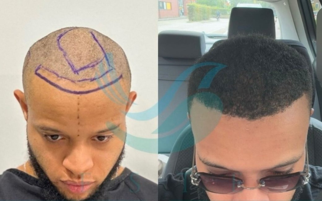
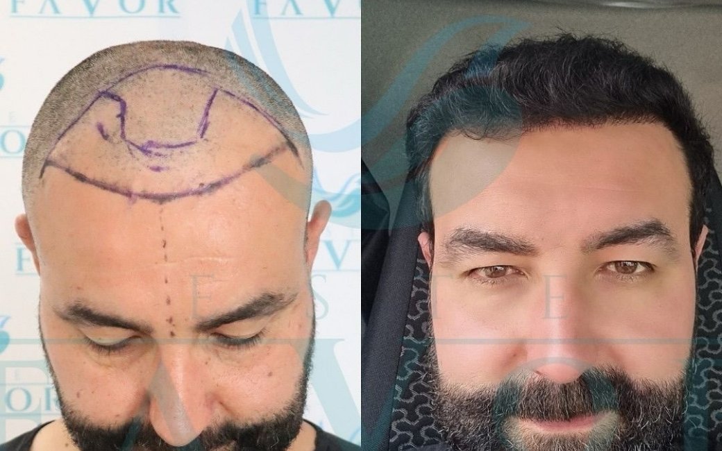
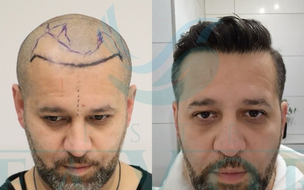
The output is a proposed hairline overlay on the patient’s photograph that can be reviewed, adjusted, and confirmed during the consultation. This serves two functions. First, it provides an objective, anatomy-based starting point for the hairline design conversation that reduces the influence of patient aesthetic preference over anatomical appropriateness. A patient who wants a very low hairline can be shown, objectively, where their facial geometry places the proportional hairline and why the proposed position is where it is. Second, it creates a documented, agreed design record that serves as a reference during the procedure itself.
The limitation of AI hairline design tools is that they operate from a fixed set of anatomical rules that do not capture all of the aesthetic judgment that an experienced surgeon applies. Hair characteristics : texture, shaft diameter, curl pattern, growth angle : all affect how a hairline will look in practice in ways that proportional geometry alone cannot fully model. The AI-generated design should be treated as a well-calibrated starting point that a surgeon refines, not as a definitive prescription.
Predictive Modelling: Projecting Pattern Progression
Some AI systems in hair restoration claim to predict the likely future progression of androgenetic alopecia based on the patient’s current presentation: their current Norwood stage, the miniaturisation distribution on trichoscopy, their age, and in some systems their reported family history. The output is a projected future pattern that is intended to inform donor allocation decisions: if the model predicts Norwood 6 at fifty, the surgical plan should allocate grafts accordingly rather than treating only the current Norwood 3 deficit.
This application of AI is the most clinically useful in concept and the most technically limited in practice. Androgenetic alopecia progression is genuinely probabilistic: there are statistical relationships between current presentation, age, family history, and eventual pattern, but the individual variance around these relationships is wide. A model trained on population data will produce a probability distribution for eventual pattern; it cannot tell any individual patient with certainty what their pattern will be at sixty.
The practical value of predictive modelling in clinical planning is not that it provides certainty but that it provides a structured, data-informed basis for the probability estimates that surgeons make anyway. An experienced surgeon examining a twenty-five-year-old with a Norwood 3 and a father who is Norwood 6 already performs an informal probabilistic assessment of where that patient is heading. An AI predictive system makes that assessment explicit, reproducible, and documentable. It does not replace clinical judgment; it gives clinical judgment a quantitative scaffold.
For patients, the practical implication is that AI-assisted prediction should be presented as a probability range rather than a definitive prognosis. A system that tells a twenty-four-year-old their pattern will reach Norwood 5 by forty is overclaiming the precision of current models. A system that tells them their trichoscopic profile and family history place them in a group where 70 percent of patients reach Norwood 5 by fifty, and that this probability estimate is one input into the graft allocation planning, is using the technology responsibly.
Where AI Cannot Replace Clinical Judgment
The consistent finding of the clinical literature on AI diagnostic tools across specialties is that they perform well within the domain they were trained on and poorly outside it. An AI trichoscopy system trained predominantly on images of male androgenetic alopecia from a specific ethnic distribution will produce less reliable results when applied to female pattern hair loss, scarring alopecia, or patients from populations underrepresented in the training data. Understanding the training domain of any AI system used in a consultation is clinically relevant.
AI systems also do not integrate information across modalities in the way a clinician does. A trichoscopy AI sees the image it is given. It does not know that the patient sitting in the chair has been losing hair rapidly for eight months, that they are under significant occupational stress, that their ferritin level came back low, or that their paternal grandfather was completely bald at thirty-five. The surgeon who reviews the AI output does know all of these things, and the synthesis of trichoscopic data with clinical history and context is what produces the diagnostic and planning accuracy that good outcomes require.
The psychological dimension of the pre-surgical assessment is entirely outside the scope of current AI tools. Whether a patient has realistic expectations, whether they show signs of body dysmorphic disorder, whether their stated goals are achievable with their available donor supply, whether they are likely to find the growth timeline psychologically manageable: none of these assessments are accessible to image analysis algorithms. A consultation that processes the AI output without addressing these dimensions is incomplete regardless of the technical sophistication of the technology it uses.
Finally, AI tools in hair restoration are only as useful as the quality of the data they receive. A poorly lit trichoscopy image, a standardised photograph taken without the correct camera-to-face distance or lighting conditions, or a donor zone dermoscopy performed on a patient whose scalp has not been properly prepared will produce AI output that is technically computed but clinically unreliable. Quality control of the imaging process upstream of the AI analysis is a human responsibility that cannot be automated.
There is also a data quality dimension to consider at the clinic level. AI systems require consistent, standardised image inputs to produce reliable outputs. A clinic that has invested in AI trichoscopy hardware but does not enforce a standardised imaging protocol, vary lighting conditions between sessions, or does not calibrate the imaging device regularly will obtain AI outputs that are technically produced but not meaningfully comparable across assessments. The availability of AI technology and the reliable use of AI technology are different things, and patients evaluating clinics should ask about the imaging protocol as well as the technology itself.

How AI Fits Into a Quality Pre-Operative Workflow
In a well-designed pre-operative workflow, AI tools occupy a specific position between initial data collection and clinical interpretation. They do not begin the assessment and they do not end it. They receive data that has been collected according to a defined protocol, produce quantified output from that data, and deliver that output to the clinician who interprets it in the context of everything else known about the patient.
The sequence in a quality consultation might look like this. The patient provides a clinical history and describes their goals. The clinician performs a visual examination and takes the relevant clinical history. Standardised trichoscopy images are captured of both the donor and recipient zones. The AI system analyses these images and produces density, miniaturisation, and shaft diameter data for each zone. The clinician reviews this data alongside the clinical history, family history, and examination findings to form a diagnostic and planning assessment. A digital hairline design is produced and reviewed with the patient. The donor zone mapping integrates the AI density data with the physical examination of the safe donor boundaries to produce the graft count estimate.’,
The output of this workflow is a surgical plan with a documented basis. The hairline position is recorded. The density targets by zone are specified. The graft count estimate is grounded in measured donor zone data rather than visual impression. The miniaturisation findings are documented as a baseline against which future assessments can be compared. This documentation also forms the basis of genuinely informed consent: the patient understands not just what the surgeon proposes to do, but why the specific design and allocation decisions were made.
For patients comparing clinics, the presence of a structured AI-integrated workflow is a meaningful quality indicator. It does not guarantee a good outcome : the quality of the surgical execution matters independently : but it indicates a clinic that is investing in the informational foundation of good planning. A clinic whose pre-hair transplant assessment consists of a visual examination and a graft count estimate is making planning decisions with substantially less information than is now available, and the results are more variable accordingly.

The Honest Appraisal
AI-assisted hair transplant planning is a genuine clinical advance, not a marketing claim. The specific capabilities it provides : rapid, reproducible, operator-independent quantification of follicular density and miniaturisation; objective facial-geometry-based hairline design proposals; structured graft count estimation from measured donor data : address real sources of variability and imprecision in the pre-operative assessment process. Clinics that have integrated these tools into a well-designed workflow are planning with better information than clinics that have not.
The honest appraisal also requires acknowledging what AI does not do: it does not replace the clinical judgment that synthesises technical data with contextual information, patient history, and psychological assessment; it does not perform the surgery; and it does not guarantee a natural result. What it does is raise the informational floor of the planning process, reducing the dependence of outcome quality on the experience of the individual clinician and creating a more consistent, documentable, reviewable basis for the decisions that determine what the patient looks like at twelve months.
