
The difference between a good hair transplant outcome and a poor one is often determined before a single graft is placed. The decisions made during the pre-operative assessment : where to place the hairline, how many grafts to harvest, which areas of the donor zone are safe to use, whether the pattern is stable or still progressing : require accurate information about the scalp at a level of detail that is not available to the naked eye. Modern clinics use a range of diagnostic technologies to gather this information systematically. Understanding what these technologies do and what they reveal is genuinely useful for any patient evaluating clinics or preparing for a consultation.
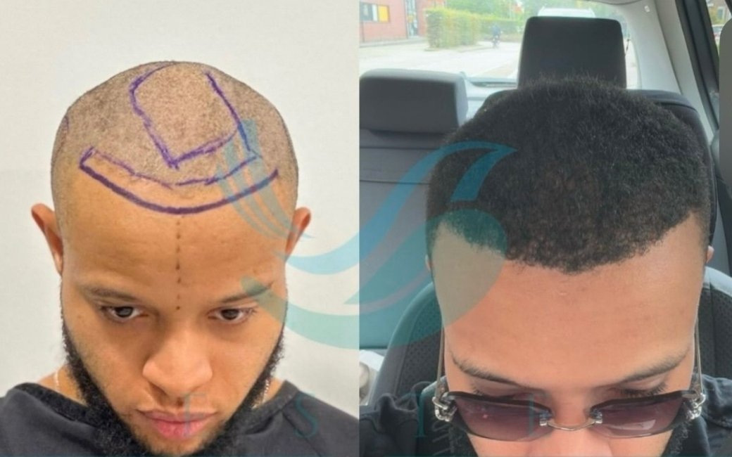
This article covers the principal scalp analysis tools used in quality hair restoration clinics: dermoscopy and trichoscopy, the phototrichogram, FUE donor zone mapping, digital hairline design tools, and the emerging role of AI-assisted analysis. For each, it explains the underlying technology, what clinical information it provides, and why that information matters for surgical planning.
Dermoscopy and Trichoscopy: The Foundation of Pre-Operative Assessment
Dermoscopy is the use of a handheld optical instrument, the dermatoscope, to examine the skin surface and sub-surface structures at magnifications typically between ten and one hundred times. The device combines a light source, a lens, and in modern versions a digital camera, allowing the examiner to see structures that are completely invisible at normal resolution. In the context of hair loss, dermoscopy reveals the architecture of individual follicles, the condition of the scalp surface, and the distribution of hair shaft diameters across different zones of the scalp.
Trichoscopy is dermoscopy applied specifically to the hair and scalp. The term was formalised by Dr. Lidia Rudnicka in the mid-2000s to describe the systematic dermoscopic examination of hair disorders. In clinical practice, trichoscopy and dermoscopy are used interchangeably when discussing scalp assessment before a hair transplant; the distinction is primarily academic.
What trichoscopy actually shows in pre-operative assessment is critical. The examiner evaluates follicular unit density across both the recipient and donor zones: how many follicular units are present per square centimetre, and how many hairs each unit contains. Crucially, it reveals follicular miniaturisation: the progressive thinning of individual hair shafts that is the hallmark of androgenetic alopecia. A follicle that is miniaturising produces a thinner, shorter hair with each growth cycle. Under trichoscopy, miniaturised hairs are visible as thin, pale, short vellus or intermediate hairs alongside terminal hairs. The proportion of miniaturised hairs in any given zone tells the surgeon how far the process has advanced there.

In the donor zone, trichoscopic assessment of miniaturisation is particularly consequential. The permanent donor zone in androgenetic alopecia is defined as the area where follicles are DHT-resistant and will not miniaturise over time. But the boundaries of this zone vary between patients, and in some patients, particularly younger ones, the lower margins of the permanent zone may be closer to the miniaturising area than classical anatomy suggests. Trichoscopy can directly confirm whether a given area of the donor zone is producing normal terminal hairs or already showing signs of miniaturisation. Grafts harvested from a miniaturising donor area will themselves progressively thin after transplantation, producing a result that deteriorates over years. Trichoscopic donor assessment is therefore not optional in quality surgical planning; it is fundamental.
The clinical significance of this is hard to overstate. A patient who is transplanted with grafts from a marginal donor area may see satisfying density in the first three to five years, followed by gradual thinning of the transplanted hairs as the miniaturisation process that was already underway in the donor follicles continues in their new location. This outcome is not caused by poor surgical technique; it is caused by inadequate pre-operative donor zone assessment. Trichoscopy prevents it.
Beyond miniaturisation, trichoscopy evaluates several other parameters relevant to surgical planning. It can identify perifollicular fibrosis: the early sign of scarring alopecia conditions that would contraindicate standard FUE. It reveals the presence of scalp inflammation, which may need to be treated before surgery. It assesses the quality of the vascular network visible at the surface, which gives indirect information about the healing capacity of the recipient zone. And it can identify the specific type of hair loss affecting the patient, distinguishing androgenetic alopecia from other conditions that may require different management.
The Phototrichogram: Quantifying Hair Density and Growth
The phototrichogram is a standardised photographic technique used to quantify hair density, hair shaft diameter, and the proportion of hairs in different phases of the growth cycle. It provides numerical data rather than qualitative assessment, making it particularly useful for tracking progression over time and for making precise surgical plans.

The basic phototrichogram protocol involves shaving a small area of the scalp, typically one square centimetre, photographing it at a defined time point, and photographing it again three to five days later. Hairs that have grown during the interval are in the anagen phase; those that have not are in telogen. By counting hair shaft diameters and growth status across the test area, the examiner can calculate the exact hair density in hairs per square centimetre, the proportion of follicles in the active growth phase, and the distribution of terminal versus vellus hair in the area.
For pre-operative hair transplant assessment, the phototrichogram is used in two main ways. First, it provides objective quantification of the donor zone density, which directly informs the graft count estimate. A donor zone with 80 hairs per square centimetre and a high proportion of multi-hair follicular units supports a different graft plan than a donor zone with 60 hairs per square centimetre, even if both look similar to the naked eye. Second, it can quantify the degree of miniaturisation in both donor and recipient zones more precisely than trichoscopy alone, because it measures actual hair shaft diameters rather than estimating them visually.
Digital phototrichograms, produced by dedicated trichoscopy systems with image analysis software, automate much of the counting and calculation process. The software identifies individual hair shafts, measures their diameters, classifies them as terminal or vellus, and calculates density figures automatically. This makes the technique faster and more reproducible than manual counting, though it requires quality imaging hardware and well-calibrated software to be reliable.
The practical implication for patients is that clinics using digital phototrichogram analysis can provide a precise graft count estimate before the procedure, not just a range. When a clinic tells you it will take between 2,000 and 3,000 grafts without having performed a formal density assessment, that range reflects uncertainty rather than precision. A measured donor zone density, combined with mapped safe extraction area, produces a graft estimate that is significantly more accurate.
Donor Zone Mapping: Planning the Harvest Before It Begins
FUE donor zone mapping is the systematic assessment of the scalp donor area to determine the safe extraction boundaries, estimate the total available graft count, and plan the distribution of extraction sites across the donor zone to avoid over-harvesting any single area.

The mapping process typically begins with a physical assessment of the safe donor zone boundaries: the posterior and lateral scalp areas that are permanently resistant to androgenetic miniaturisation. These boundaries are identified based on clinical observation, family history, and trichoscopic assessment of the peripheral donor zone. In a well-conducted assessment, the surgeon marks or mentally notes the zones of confirmed stability and identifies the transitional margins where stable and unstable areas meet.
Within the mapped donor zone, the density assessment determines how many grafts can be safely harvested. The standard safe extraction density for FUE is generally 10 to 15 grafts per square centimetre; above this density, visible thinning of the donor area occurs. By calculating the surface area of the safe donor zone and the density of follicular units within it, the surgeon can estimate the available graft count across one or multiple sessions. This calculation is directly relevant to the planning conversation at twenty-five versus the planning conversation at fifty-five: the lifetime donor budget is a defined number, and it must be allocated intelligently.
Advanced clinics use digital mapping systems, typically tablet-based software or dedicated devices, that allow the surgeon to mark the donor zone boundaries, record density measurements in different sub-regions, and calculate the available graft count interactively. Some systems integrate with the trichoscopic assessment, automatically importing density data from the phototrichogram into the donor zone map. The output is a visual representation of the donor zone that can be reviewed with the patient during consultation, making the graft planning process transparent rather than opaque.
For patients, the most immediately useful output of donor zone mapping is the lifetime graft budget estimate. When a surgeon tells a twenty-five-year-old that their donor zone contains approximately 5,500 harvestable grafts across their lifetime, and then shows them what Norwood 4 and Norwood 5 coverage requires in graft numbers, the conversation about conservative planning is grounded in specific arithmetic rather than abstract advice. This transforms the quality of the planning conversation.
Digital Hairline Design: Visualising the Result Before Surgery
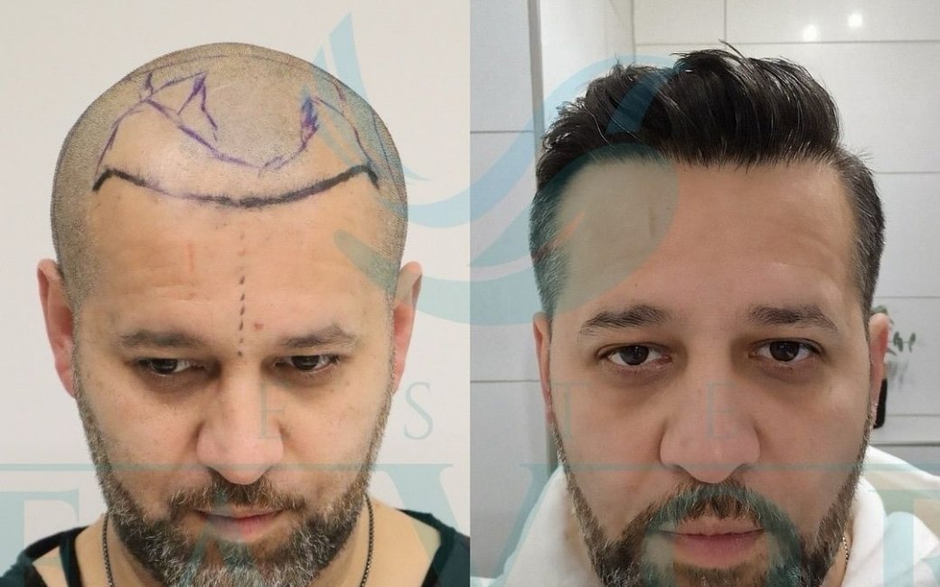
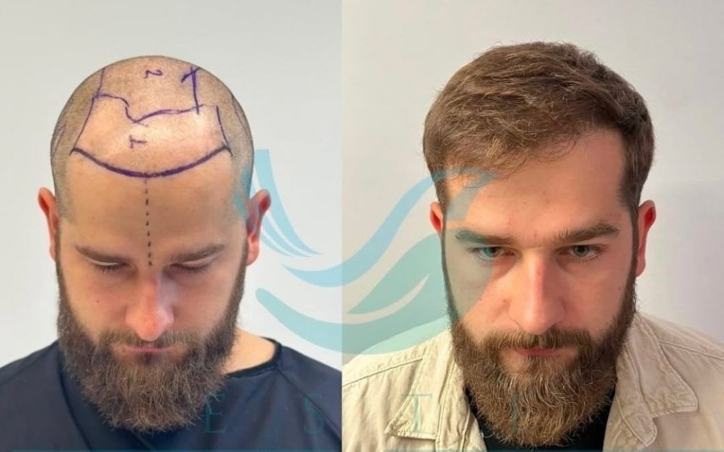
Digital hairline design tools allow the surgeon and patient to visualise the planned hairline position on the patient’s actual face before any grafts are placed. The technology ranges from simple photo-editing overlays to dedicated software platforms that model three-dimensional scalp geometry and allow hairline design in multiple orientations.
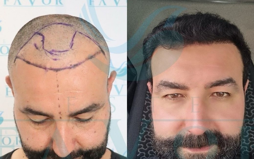
At its most basic level, digital hairline design involves marking the planned hairline position on a high-resolution photograph of the patient’s scalp. The surgeon draws the proposed hairline using the software, considering the patient’s facial proportions, the natural hairline characteristics appropriate for their age and sex, and the available graft supply. The patient can then review the design, and adjustments can be made iteratively before a commitment is made.
More sophisticated systems use standardised facial landmark analysis to generate a proposed hairline position based on the patient’s facial geometry. The software identifies the mid-facial point, the brow line, and the existing hairline boundary, and uses these reference points to calculate a proportional hairline position. This provides a quantitative basis for the design conversation rather than relying entirely on the surgeon’s visual judgment.
The value of digital hairline design extends beyond the aesthetics of the result. It creates a documented record of what was planned and agreed upon before surgery. It allows the patient to evaluate a conservative design versus a more aggressive design and understand the visual difference before committing. And it gives the surgeon a working template for the procedure itself: during the operation, the planned hairline can be referenced and the graft placement guided accordingly.
The documentation function of digital hairline design is also relevant from a patient protection standpoint. A signed, dated record of the agreed hairline position protects both parties if there is any subsequent disagreement about what was planned. In quality clinics, this documentation is part of the standard informed consent process.
AI-Assisted Scalp Analysis: What It Can and Cannot Do
Artificial intelligence tools for scalp and hair analysis have entered clinical practice in recent years, primarily as analysis engines integrated with digital trichoscopy hardware. These systems use convolutional neural networks trained on large datasets of annotated scalp images to classify hair loss patterns, measure follicular density, quantify miniaturisation, and in some systems predict the likely progression of androgenetic alopecia based on the patient’s current presentation.
The genuine clinical value of AI scalp analysis lies in its speed and reproducibility. A trained AI system can analyse a trichoscopic image and produce quantified density, miniaturisation percentage, and hair shaft diameter distribution in seconds, with consistent results across different operators and different sessions. This removes the inter-observer variability that affects manual trichoscopic assessment and provides a standardised baseline for tracking change over time.
Where AI scalp analysis is currently less reliable is in contextual clinical judgment. The AI system knows what it sees in the image; it does not know the patient’s age, family history, rate of progression over the past three years, or the broader clinical context that an experienced surgeon integrates into every assessment. A miniaturisation percentage of thirty in the donor zone means something different in a twenty-three-year-old than in a fifty-year-old. The AI does not make that distinction without explicit input; the surgeon must.
That said, the integration of AI analysis into systematic pre-operative workflows is one of the genuine advances in hair restoration practice over the past decade. It means that the quality of the trichoscopic information available at consultation no longer depends entirely on the skill and experience of the individual examiner. A well-trained AI system with a calibrated camera can extract consistent, reproducible density and miniaturisation data from any scalp, providing a reliable baseline regardless of operator variability.
The responsible position on AI scalp analysis is that it is a genuinely useful tool that enhances and standardises the information available to the surgeon, not a replacement for clinical judgment. Clinics that use AI analysis should be using it to supplement a thorough manual assessment, not to substitute for it. The patient whose scalp has been analysed by a well-calibrated AI trichoscopy system and reviewed by an experienced surgeon is in a better position than a patient whose assessment relied on either alone.
How These Technologies Work Together in Clinical Practice
In a quality pre-operative assessment, these technologies are not used in isolation but as a coordinated workflow that builds from initial observation to detailed quantification to surgical planning. The sequence typically looks like this.

The consultation begins with a clinical history and visual examination, gathering information about the rate and pattern of hair loss, family history, any relevant medical conditions, and the patient’s goals. This establishes the clinical context within which the technical assessment is interpreted.
Trichoscopic examination of the recipient and donor zones follows, providing qualitative and semi-quantitative information about miniaturisation, follicular density, and scalp health. In many clinics this is combined with AI-assisted analysis to produce quantified metrics alongside the surgeon’s clinical observations.
Where indicated, phototrichogram assessment provides precise numerical density data for the donor zone and confirms the miniaturisation distribution. This step is particularly important for patients where the donor zone assessment is ambiguous or where the graft plan requires a precise donor count.
Donor zone mapping integrates the density data with the safe extraction zone boundaries to produce the graft plan: the total available grafts, the session allocation, and the extraction distribution across the donor area. Digital hairline design then establishes where the grafts will go in the recipient zone, providing a visual representation of the intended result that the patient can review before committing to the procedure.
The output of this workflow is a surgical plan based on objective data rather than clinical impression alone. It is documentable, reviewable, and revisable. It gives the patient a transparent basis for informed consent. And it gives the surgeon the information needed to make consistently good decisions about graft allocation, hairline design, and the overall architecture of the result.
For patients comparing clinics, the presence or absence of systematic pre-operative technology use is one of the more reliable quality signals available. A clinic that uses dermoscopy, has a documented donor zone assessment process, and shows patients their planned hairline in a digital format before the procedure is demonstrating a commitment to precision planning that is not universal in the market. It is not a guarantee of a good result, but it is a meaningful indicator of a clinic that treats the planning phase with the seriousness it deserves.
What to Expect at a Quality Consultation
Patients attending a consultation at a quality hair restoration clinic should expect to see some combination of the technologies described in this article in use. A clinic that bases its graft count estimate and hairline design on visual inspection alone, without any magnified assessment of the donor or recipient zone, is working with less information than is available and making decisions with less precision than the current standard of care supports.
The specific combination of tools used will vary by clinic and by the complexity of the individual case. Not every patient needs a full phototrichogram or an AI-assisted analysis. But every patient should have a magnified assessment of the donor zone to confirm its quality before grafts are planned, and every patient should have a documented record of the planned hairline position before the procedure begins. These are not luxury features of sophisticated clinics; they are the baseline of responsible surgical planning.
Technology is a means, not an end. The best pre-operative assessment combines the objective data from modern diagnostic tools with the clinical judgment of an experienced surgeon who understands what the numbers mean in the context of the individual patient. That combination, applied consistently across every consultation, is what separates clinics that produce reliably good outcomes from those that produce variable ones.
